


 Foundational characteristics of cancer include proliferation, angiogenesis, migration, evasion of apoptosis, and cellular immortality. Find key markers for these cellular processes and antibodies to detect them.
 The SUMOplot™ Analysis Program predicts and scores sumoylation sites in your protein. SUMOylation is a post-translational modification involved in various cellular processes, such as nuclear-cytosolic transport, transcriptional regulation, apoptosis, protein stability, response to stress, and progression through the cell cycle.
 The Autophagy Receptor Motif Plotter predicts and scores autophagy receptor binding sites in your protein. Identifying proteins connected to this pathway is critical to understanding the role of autophagy in physiological as well as pathological processes such as development, differentiation, neurodegenerative diseases, stress, infection, and cancer.
> home > Products > Primary Antibodies > Antibody Collections > Goat Antibodies > Histamine Receptor H2 (aa309-323) Antibody (internal region)
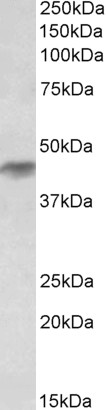
Histamine Receptor H2 (aa309-323) Antibody (internal region)
Peptide-affinity purified goat antibody
- SPECIFICATION
- CITATIONS: 1
- PROTOCOLS
- BACKGROUND

Application
| WB, E |
|---|---|
| Primary Accession | P25021 |
| Other Accession | NP_001124527.1, NP_071640.1, 3274 |
| Reactivity | Human |
| Host | Goat |
| Clonality | Polyclonal |
| Concentration | 0.5 mg/ml |
| Isotype | IgG |
| Calculated MW | 40098 Da |
| Gene ID | 3274 |
|---|---|
| Other Names | Histamine H2 receptor, H2R, HH2R, Gastric receptor I, HRH2 |
| Dilution | WB~~1:1000 E~~N/A |
| Format | 0.5 mg/ml in Tris saline, 0.02% sodium azide, pH7.3 with 0.5% bovine serum albumin |
| Storage | Maintain refrigerated at 2-8°C for up to 6 months. For long term storage store at -20°C in small aliquots to prevent freeze-thaw cycles. |
| Precautions | Histamine Receptor H2 (aa309-323) Antibody (internal region) is for research use only and not for use in diagnostic or therapeutic procedures. |
| Name | HRH2 |
|---|---|
| Function | G-protein coupled receptor for histamine, primarily mediating gastric acid secretion. Predominantly expressed in the gastric mucosa, couples to G(s) G alpha proteins upon histamine binding, leading to activation of adenylate cyclase and increased intracellular cyclic AMP (cAMP) levels (PubMed:38647423, PubMed:39333117). This signaling cascade stimulates parietal cells to secrete hydrochloric acid, playing a key role in digestive physiology. Also expressed in other tissues, including the heart and central nervous system, where it may contribute to cardiac stimulation and modulate neurotransmitter release (By similarity). |
| Cellular Location | Cell membrane; Multi-pass membrane protein. |
Research Areas
Citations ( 0 )

Application Protocols
Provided below are standard protocols that you may find useful for product applications.
Background
This antibody is expected to recognize both reported isoforms (NP_001124527.1; NP_071640.1).
References
Histamine: its novel role as an endogenous regulator of Con A-dependent T cell proliferation. Nakane H, Sonobe Y, Watanabe T, Nakano K. Inflamm Res. 2004 Jul;53(7):324-8. PMID: 15241568
Abcepta welcomes feedback from its customers.
If you have used an Abcepta product and would like to share how it has performed, please click on the "Submit Review" button and provide the requested information. Our staff will examine and post your review and contact you if needed.
If you have any additional inquiries please email technical services at tech@abcepta.com.
$ 423.00
Cat# AF3587a
Ordering Information
United States
AlbaniaAustraliaAustriaBelgiumBosnia & HerzegovinaBrazilBulgariaCanadaCentral AmericaChinaCroatiaCyprusCzech RepublicDenmarkEstoniaFinlandFranceGermanyGreeceHong KongHungaryIcelandIndiaIndonesiaIrelandIsraelItalyJapanLatviaLithuaniaLuxembourgMacedoniaMalaysiaMaltaMexicoNetherlandsNew ZealandNorwayPakistanPolandPortugalRomaniaSerbiaSingaporeSlovakiaSloveniaSouth AfricaSouth KoreaSpainSwedenSwitzerlandTaiwanTurkeyUnited KingdomUnited StatesVietnamWorldwideOthers
USA Headquarters
(888) 735-7227 / (858) 622-0099 or (858) 875-1900
Other Products
Shipping Information
Domestic orders (in stock items)
Shipped out the same day. Orders placed after 1 PM (PST) will ship out the next business day.
International orders
Contact your local distributors
